Maka Medical Surgical
Choosing the right Intramedullary Nail in 2026 requires more than comparing product brochures. It requires careful judgment, current evidence, and practical surgical experience. The best implant depends on fracture location, bone quality, canal anatomy, and the patient’s functional needs. A nail that performs well in a femoral shaft fracture may be unsuitable for a complex tibial injury.
This guide examines the features that matter during real clinical decision-making. These include nail diameter, length, curvature, locking options, insertion technique, material, and instrument reliability. It also considers evidence from peer-reviewed research, manufacturer documentation, and established orthopedic practice. Imaging remains essential. A three-dimensional understanding of the medullary canal can prevent avoidable sizing errors before surgery.
Small details matter.
For example, a difficult locking screw trajectory may increase operative time and frustration. A poorly matched curvature may create stress at the entry point. Experienced surgeons also consider soft-tissue condition, fracture pattern, reduction quality, and the availability of compatible tools. Device quality alone cannot replace sound planning.
No checklist is perfect. Clinical decisions still involve uncertainty, especially when evidence is limited for newer designs. Some promising features may lack long-term clinical validation. Therefore, readers should compare published outcomes with local experience and applicable regulatory requirements. Consultation with qualified orthopedic specialists remains essential.
By weighing biomechanics, usability, patient-specific anatomy, and reliable clinical evidence, this article aims to support a more disciplined choice. The goal is not to promote one brand. It is to help clinicians ask better questions before selecting an Intramedullary Nail for 2026 and beyond.
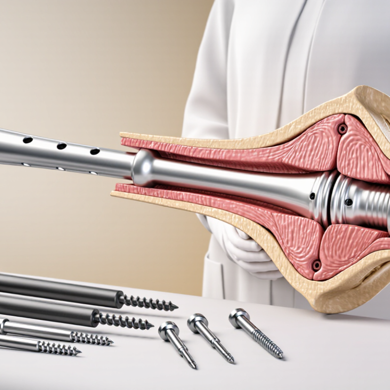
Choosing an intramedullary nail starts with defining the fracture, not selecting a device by reputation. Is the break in the femur, tibia, or humerus? Is it near a joint, in the shaft, or close to the bone’s end? A simple transverse fracture and a segmental fracture place different demands on fixation. Details matter. Review X-rays from more than one angle, and consider CT when the fracture pattern is unclear.
The nail’s length, diameter, curvature, and locking options should match the patient’s anatomy and the fracture’s location. For example, a very proximal or distal fracture may need careful attention to how locking screws control alignment. Bone quality, canal shape, soft-tissue condition, and other injuries also influence the plan. The treatment goal may be stable alignment that allows early movement, but the right balance depends on the individual case. A clean-looking image can still leave questions about stability or hidden fragments; that uncertainty deserves discussion, not guesswork. The treating orthopedic surgeon should compare imaging, examination findings, and the patient’s recovery needs before choosing a nail.
| Fracture pattern | Typical location | Nail features to consider | Treatment goals | Planning checks |
|---|---|---|---|---|
| Femoral shaft fracture Transverse, short-oblique, or selected comminuted patterns |
Mid-shaft femur | A reamed or unreamed locked nail may be considered. Choose length and diameter to fit the femoral canal; locking options help control length and rotation. | Restore alignment, length, and rotation; provide stable fixation that supports mobilization and fracture healing. | Assess fracture extension, canal dimensions, existing implants, and whether the fracture can be reduced and controlled with a nail. |
| Subtrochanteric fracture Often unstable, with a tendency to deform |
Proximal femur, just below the lesser trochanter | A long cephalomedullary-style nail with proximal fixation into the femoral head and neck may be considered, depending on the fracture and anatomy. | Control varus and rotational deformity, maintain alignment, and stabilize the proximal and shaft fragments. | Check fracture extension, reduction quality, bone quality, femoral-head fixation options, and the planned nail entry point. |
| Intertrochanteric fracture Stable or unstable patterns |
Proximal femur between the greater and lesser trochanters | A short or long cephalomedullary nail may be selected according to fracture extension, stability, and associated shaft involvement. Proximal fixation is designed to support the head-neck fragment. | Maintain reduction and limit collapse or rotation while enabling an appropriate rehabilitation plan. | Evaluate lateral-wall integrity, reverse-obliquity or subtrochanteric extension, bone quality, and reduction on imaging. |
| Distal femur fracture Selected extra-articular or metaphyseal patterns |
Lower femur, above the knee | A retrograde nail with suitable distal interlocking options may be considered. Very distal or complex articular fractures may require another fixation strategy or combined fixation. | Restore limb alignment and length, secure the distal segment, and preserve knee function where possible. | Confirm distal fragment size, joint-surface involvement, knee access considerations, and compatibility with any hip or knee implant. |
| Tibial shaft fracture Transverse, oblique, or selected comminuted patterns |
Mid-shaft tibia | A locked tibial nail is commonly considered. Nail length, diameter, and proximal and distal locking options should match the canal and fracture configuration. | Maintain alignment and rotational control while providing stable fixation for progressive rehabilitation when appropriate. | Assess soft-tissue injury, open-fracture status, canal anatomy, fibular injury, and the ability to obtain secure fixation on both sides of the fracture. |
| Proximal or distal tibial fracture Metaphyseal pattern or short segment |
Near the knee or ankle | A nail with multiple interlocking options near the fracture may help control a short segment. A plate or another method may be preferable when alignment or articular reconstruction cannot be reliably achieved with a nail alone. | Achieve and maintain alignment, especially in the coronal and sagittal planes, and protect any joint-surface reconstruction. | Review the fracture’s distance from the joint, articular involvement, soft tissues, and the available space for interlocking screws. |
| Humeral shaft fracture Selected closed or open shaft patterns |
Mid-shaft humerus | A humeral nail may be considered in selected cases. Entry approach, locking configuration, and proximity to the shoulder or elbow are important; plating is also commonly used. | Stabilize the shaft while accounting for shoulder and elbow motion and the individual’s functional needs. | Assess fracture location, radial-nerve status, bone quality, shoulder condition, and whether the entry route is suitable. |
Selection depends on imaging, fracture stability and extension, bone and canal anatomy, soft-tissue condition, existing implants, and patient-specific risks. This table is a general educational guide, not a substitute for evaluation and treatment planning by a qualified orthopedic surgeon.
Start with the bone and fracture pattern, not the device catalog. Identify the femur, tibia, or humerus. Note whether the fracture is near a joint or within the shaft. A simple transverse break needs different control than a segmental fracture. Details matter.
Review full-length X-rays from multiple angles. Use CT when fragments or fracture lines remain unclear. Compare imaging with physical examination findings. A clean image may still hide instability. Do not replace discussion with guesswork.
Measure the canal at its narrowest point. A wide nail may increase insertion pressure. A small nail may provide limited stability. Match length, diameter, and curvature to the bone. The nail should avoid joint penetration and excessive prominence.
Locking choices should reflect the fracture location. Proximal fractures may need several multidirectional screws. Distal fractures need secure fixation near the joint. Shaft fractures may require a balanced design instead. More features are not always better.
Bone quality, canal shape, soft-tissue condition, and other injuries influence the plan. Entry-point access also matters. Reaming technique and reduction quality can change the final result. Anatomy can be surprisingly irregular.
Review studies, trauma registries, and long-term follow-up results. Check union, alignment, infection, reoperation, and functional recovery. Ask whether the evidence includes similar fractures. Evidence is rarely perfect.
Yes. Familiarity with instruments and reduction methods can reduce avoidable handling problems. A theoretically stronger option may be less safe in unfamiliar hands. Training helps, but judgment develops unevenly. That limitation deserves honesty.
Discuss entry-point risks, rotational control, screw lengths, and possible later procedures. Confirm the canal shape, fracture level, and available instruments. Document why the chosen dimensions fit the anatomy. This can still be wrong, so reassessment matters.
Assess safety before incision, during implantation, and throughout follow-up. Watch alignment during reduction, not only after fixation. Review healing and function over time. Small problems can grow.
Choosing the best Intramedullary Nail in 2026 begins with a clear understanding of the fracture, including its type, location, and the patient’s treatment goals. The nail’s length, diameter, shape, and configuration should suit the bone’s anatomy and the fracture pattern, while allowing the surgeon to achieve appropriate alignment and stability. Material properties, locking options, and compatibility with available instruments are also important considerations because they can affect surgical planning and fixation.
Before making a selection, review relevant clinical evidence and safety information, and consider the experience of the surgical team with the proposed technique. Confirm that the device is appropriately approved and available, and that long-term support, training, and follow-up resources can be accessed. A careful, case-specific comparison helps balance practical needs with patient factors and supports an informed treatment decision.